Arthroplasty imaging · Core lab · RSA · CT-RSA

Imaging, core lab and RSA for arthroplasty studies

Radiographs are collected in most arthroplasty studies, but collecting images is not the same as generating dependable imaging evidence. Differences in positioning, projection, equipment, timing and reader interpretation can make an apparently simple radiographic endpoint impossible to compare across sites.

Imaging charterIndependent core labRSA & CT-RSAMigration & fixationEU MDR 2017/745
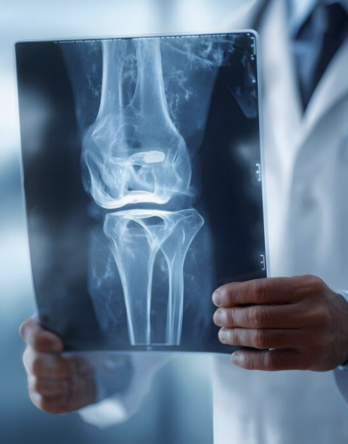
Knee radiograph on a viewing box, central imaging review for arthroplasty studies
From device claim to imaging conclusion
1
Device innovation & residual uncertainty
2
Imaging question
3
Modality & measurement method
4
Timepoint & interpretation rule
5
Independent assessment
6
CSR, PMCF & CER
The modality should be chosen because it answers the clinical question, not because it is routinely available.
Trusted by

Leading medical device teams

TERUMOMerilNIHON KOHDENVYGONColoplastSHOFUASAHI INTECCRegenLabTERUMOMerilNIHON KOHDENVYGONColoplastSHOFUASAHI INTECCRegenLab
Who oversees the imaging

Orthopedic clinical oversight on every imaging endpoint

EUCROF Platinum Award 2026
EUCROF Platinum Award 2026xShare Open Call for Clinical Research, co-funded by the European Union
Dr Nikhil Khadabadi

Dr Nikhil Khadabadi

CMO · Orthopedics & Spine
NHS orthopedic surgeon

Former reviewer atTUV SUD
20+yrs

in orthopedic surgery & Class III implant evidence

  • Assesses Class III orthopedic & spinal evidence under EU MDR
  • Former Principal Investigator, Stryker robotic surgery trial
  • Leads CERs, PMCF & registries for Orthopedics & Spine
LinkedIn
Dr Mark Da Costa

Dr Mark Da Costa

Chief Operating Officer · former TÜV SÜD Senior Reviewer · Senior Consultant Surgeon

25+yrs

in device evaluation & Notified Body review

  • Assessed 400+ medical devices in Europe
  • Brings the reviewer perspective to protocol & CER design
  • Oversees delivery across the full evidence program
LinkedIn
Sébastien Meier Piantanida

Sébastien Meier Piantanida

Chief Data Officer · Biometrics & Data Systems

30yrs

in clinical data management, EDC & biometrics

  • Leads EDC, data management & biostatistics
  • Expertise in CDISC, CDASH, SDTM & ADaM
  • Validated data flows between sites, core lab & statistics
LinkedIn
Who this page is for

When imaging has to carry evidential weight

This page is for teams whose images must support a conclusion, not simply document the procedure.

A fixation, migration or alignment claim

The implant makes a claim that only imaging can substantiate.

Radiographic performance endpoints

The protocol includes endpoints derived from images.

Images across several sites or countries

Comparability between centers becomes the limiting factor.

Centers using different acquisition methods

Variation in positioning and projection is already present.

Independent review is required

The sponsor needs assessment separated from site interpretation.

Radiolucent lines or loosening are outcomes

Findings that are notoriously inconsistently defined.

RSA is being considered

An early study where migration may be the central question.

Imaging must support PMCF or a CER update

Post-market evidence that has to withstand review.

« Radiographs will be assessed »

The protocol says imaging will happen, without defining how.

The objectivity has been questioned

A Notified Body or authority has challenged the imaging evidence.

Clinical evidence for arthroplasty medical devices — the parent hub. For the clinical side of the endpoint logic, see arthroplasty endpoints and PROMs.

Claim first

The imaging question must follow the device claim

Imaging strategy follows the same decision logic as endpoint strategy. The claim and the residual uncertainty define the imaging question; only then do modality, measurement method, timepoint and interpretation rule follow.

Device innovationRemaining uncertaintyImaging questionModalityMeasurement methodTimepointInterpretation ruleClinical & regulatory conclusion

New cementless coating

Is early fixation developing as expected?
Standard radiographs, RSA or CT-RSA.

New acetabular component

Is cup position within the intended range?
Standardized pelvic radiographs or CT.

New knee alignment philosophy

Is the planned component alignment achieved?
Long-leg and joint-specific radiographs.

New glenoid component

Is fixation maintained over follow-up?
Standardized shoulder radiographs, or CT where justified.

Patient-specific implant

Do fit and position match the preoperative plan?
CT-based planning and postoperative imaging.

Robotic implantation system

How closely does achieved position match the plan?
System data combined with radiographic or CT confirmation.

Definitions

Imaging endpoint, measurement and interpretation are different things

As with clinical outcomes, precision of language is what prevents a weak imaging endpoint.

Imaging domain

What is assessed: position, alignment, fixation, migration, radiolucency, osteolysis, loosening or implant integrity.

Measurement method

How it is assessed: an angle, a distance, a zone-based score, presence or absence, change over time, three-dimensional translation or rotation, or composite radiographic criteria.

Imaging endpoint

A prespecified result, for example the change in femoral stem subsidence from baseline to twenty-four months.

Interpretation criterion

The rule used to classify a finding as stable, progressive, clinically significant, indeterminate or unevaluable.

Key message. Radiographic assessment is not a complete endpoint. The view, the measurement, the reader, the timepoint and the interpretation rule must all be prespecified.

Three levels of evidence

Use the lowest level that answers the question reliably

Arthroplasty imaging is not a single service. Three connected levels answer different questions at different levels of cost and complexity.

Level 1 · Standardized site imaging

Images acquired at clinical sites under a common imaging charter. Suitable for routine assessment of position and alignment, monitoring of radiolucent lines, fracture or dislocation, implant integrity and standard clinical follow-up.

Level 2 · Independent core-lab assessment

Central review by trained independent readers against prespecified criteria. Suitable when consistency across centers, blinded review, quantitative measurement, longitudinal comparison or regulatory-grade evidence is required.

Level 3 · RSA or CT-RSA

Specialized measurement of three-dimensional implant migration relative to bone. Suitable for new fixation concepts, cementless implants, new coatings or porous structures, and small technically intensive early studies.

Traditional marker-based RSA uses simultaneous radiographs and a calibration cage to quantify implant or skeletal movement in three dimensions. CT-RSA is an evolving alternative that uses serial CT data rather than the conventional biplanar marker-based workflow.

Biplanar hip radiographs used to compare imaging modalities in arthroplasty
A more complex modality is not automatically strongerThe best method is the one that answers the question with proportionate burden and acceptable precision.
Three connected levels

The level follows the question

Arthroplasty imaging is not a single service. Three connected levels answer different questions at different levels of cost and complexity.

Three connected levelsA more complex modality is not automatically strongerLevel 1Standardized site imagingImages acquired at clinical sitesunder a common imaging charter.Position and alignmentRadiolucent linesFracture or dislocationImplant integrityRoutineLevel 2Independent core-lab assessmentCentral review by trained independentreaders against prespecified criteria.Consistency across centersBlinded reviewQuantitative measurementRegulatory-grade evidenceCentralLevel 3RSA or CT-RSASpecialized measurement of three-dimensionalimplant migration relative to bone.New fixation conceptsCementless implantsNew coatings, porous structuresSmall early studies3DThe level follows the question that has to be answered, and the cost and complexity that question justifies.
Site imaging, independent core lab, RSA and CT-RSA.
Modality selection

What each modality answers well, and what it does not

  • Plain radiography — position, alignment, radiolucency, fracture, loosening, implant integrity. Watch: positioning, projection and reader variability.
  • Long-leg radiography — limb and component alignment in knee studies. Watch: reproducible stance and rotational positioning.
  • CT — three-dimensional position, osteolysis, component orientation, bone assessment. Watch: radiation, metal artifact, cross-site protocol consistency.
  • EOS or low-dose biplanar imaging — alignment and functional position where available. Watch: access, calibration and comparability.
  • Conventional RSA — highly precise implant-migration measurement. Watch: specialist equipment, markers, cage and trained sites.
  • Model-based RSA — migration without attaching markers to the implant. Watch: availability of validated implant models and software.
  • CT-RSA — three-dimensional migration from serial CT. Watch: protocol, dose, segmentation, validation and site capability.
  • MRI — selected soft-tissue or adverse local tissue reaction questions. Watch: implant compatibility and metal artifact.
  • Ultrasound — selected soft-tissue or fluid assessments. Watch: operator dependence, limited implant position assessment.
Imaging charter

The charter is the operational foundation

The imaging charter is what turns an imaging intention into a repeatable process. It defines objectives, modality, required views and projections, patient positioning, side and anatomical coverage, equipment, acquisition parameters, calibration, the permitted acquisition window and the baseline definition — then file format, naming and de-identification, transfer, quality-control criteria, repeat-image rules, reading methodology, reader blinding, measurement definitions, adjudication rules, handling of missing or unevaluable images, and version control.

Core labFrom acquisition at the site to an analysis-ready endpoint1Site acquisitionPosture, projection,limb rotation, equipment2Transfer &de-identificationFile format, naming,controlled transfer3Quality controlAcceptance criteria,repeat-image rules4Blinded readingTrained readers, definedmeasurement rules5AdjudicationReader disagreement resolvedby prespecified rule6Analysis-ready dataReconciled with the EDC,version controlledControl points written into the imaging charterNon-evaluable imagesReader disagreementMissing timepointsMeasurement driftVersion controlA core lab is an operational system, not a single reader.
Core lab workflow, from acquisition at the site to analysis-ready data.
Acquisition & baseline

Consistency begins at the imaging site

Standardized acquisition

  • Patient posture; weight-bearing or non-weight-bearing
  • Limb rotation, beam direction, source-to-image distance
  • Joint-centered versus long-leg imaging, required landmarks
  • Magnification marker where applicable, exposure and resolution
  • Timing relative to surgery; same modality at follow-up
  • Bilateral or unilateral acquisition; documentation of deviations

Baseline — the reference image

  • Preoperative or postoperative reference, and the permitted window
  • Whether weight-bearing is allowed; imaging before mobilization
  • How incomplete baseline images are handled
  • Whether the reference must pass central quality control
  • How later images are matched to it
  • For conventional RSA, a clearly defined early postoperative reference examination

Small differences in positioning alter apparent alignment, component orientation, joint geometry and interface visibility. A technically correct but non-standardized image can be clinically useful and still be unsuitable for a quantitative endpoint. In practice, an illustrated acquisition manual is one of the highest-value documents in the study: it shows acceptable images, common positioning errors, correct landmark coverage, images requiring repeat acquisition, and images that remain clinically usable but are not endpoint-evaluable.

Important. The imaging charter should be finalized before site activation and reflected in the protocol, the CRF, the monitoring plan, the data-management plan and the statistical analysis plan.

Component position & alignment

The measures are joint-specific

The lists below are illustrative rather than prescriptive: the selection should follow the claim and the residual risk.

Hip

Acetabular inclination and anteversion, femoral stem alignment, femoral offset, leg-length difference, center of rotation, stem subsidence, cup migration, component seating, fracture, and component position in relation to dislocation.

Knee

Mechanical or anatomical limb alignment, femoral and tibial coronal alignment, femoral flexion, tibial slope, component rotation where CT is used, joint-line position, patellar position and tracking, component overhang, implant seating, planned versus achieved alignment.

Shoulder

Humeral component position, glenoid inclination and version, component seating, scapular notching, tuberosity healing, acromial or scapular spine fracture, radiolucency, migration or loosening, implant integrity.

Other joints

Ankle component alignment, elbow loosening and implant integrity, small-joint implant migration or failure, and joint-specific radiographic classification systems where the device and its claims require them.

Planned versus achieved

Robotics, navigation and patient-specific implants

Particularly relevant for robotic arthroplasty, navigation-assisted implantation, patient-specific instruments and implants, 3D-printed components and preoperative CT-based planning systems.

  • Which preoperative plan is the reference
  • Whether software data or independent imaging is the definitive source
  • How coordinate systems are aligned
  • The tolerance limits
  • Whether absolute deviation or categorical accuracy is used
  • Which patients are excluded from the positional analysis
  • How intraoperative plan changes or conversion to manual instrumentation are managed

Key message. Data generated by the surgical system should not automatically be treated as independent confirmation of that system’s accuracy.

Radiolucent lines, loosening & osteolysis

The findings most often reported and least often defined

Radiolucent lines — what to define

  • The anatomical zones and the interface being assessed
  • Width and extent; complete or incomplete
  • Immediate or newly developed; stable or progressive
  • Presence of symptoms; relationship to migration or loosening
  • Reader confidence and image-evaluability criteria
  • Report: patient-level and component-level prevalence, zone distribution, width or extent, progression, association with symptoms, migration, reoperation or revision

Four terms that are not synonyms

  • Radiographic loosening — a prespecified combination of findings such as progressive migration, interface change or component displacement
  • Clinical loosening — radiographic findings together with pain, instability, loss of function or revision findings
  • Osteolysis — a focal or progressive area of bone loss assessed against predefined imaging criteria
  • Implant failure — fracture, dissociation, wear, migration, loss of fixation, revision or planned revision

Important clinical distinction. A radiolucent line is a radiographic finding, not automatically proof of osteolysis or mechanical loosening. Early lines may relate to implantation technique, cement penetration, component position or interface characteristics, and their meaning depends on timing, progression and accompanying findings. Do not use loosening, radiolucency, osteolysis and revision as interchangeable terms.

Multicenter imaging

One operational imaging model across the sites

Multicenter imaging only works when every center is qualified, trained and monitored against the same charter. Equipment, DICOM capability and local radiation review differ market by market.

ARTHROPLASTY IMAGING & RSA NETWORK - DACH ARTHROPLASTY IMAGING & RSA NETWORK · DACH DEATCH Site qualificationModality, equipment model, DICOM capability,calibration kit and transfer routeWatch: test-image approval before thefirst patientGermanyHigh-volume centers, established researchimaging and RSA experienceWatch: local radiation review timelinesAustriaSpecialist centers, consistent equipmentand technologist availabilitySwitzerlandStrong imaging infrastructure; SIRIS forlong-term follow-upWatch: separate Swiss pathway andcross-site CT calibration

Imaging capability, calibration equipment and radiation approvals are assessed center by center during feasibility.

Longitudinal assessment

A single image describes a moment; fixation evidence comes from behavior over time

The strategy should distinguish new findings, persistent findings, progressive findings, resolved findings, findings present at baseline, findings appearing after an intercurrent event, and findings assessed after revision or reoperation.

Longitudinal endpoints

  • Proportion of implants with progressive radiolucent lines at twenty-four months
  • Mean change in stem subsidence from baseline
  • Proportion of components remaining radiographically stable
  • Time to first radiographic sign of loosening
  • Change in component position
  • Association between early migration and later clinical outcomes

Imaging timepoints

  • Preoperative — anatomy, deformity and planning
  • Immediate postoperative — reference position and procedural result
  • Six weeks — early position, fracture or displacement
  • Three months — early fixation or migration pattern
  • Six months — stabilization or continued migration
  • Twelve months — position, fixation and radiographic progression
  • Twenty-four months — established migration and interface findings
  • Longer term — wear, osteolysis, loosening and survivorship

Key message. RSA studies may need more frequent early assessments than routine radiographic PMCF studies. The schedule should follow the process being measured, not convention.

Independent assessment

A core lab is an operational system, not a single reader

Independent reading means more than sending images to a radiologist. A core laboratory provides imaging-charter development, site and equipment qualification, acquisition training, central image receipt, de-identification checks, image-quality review and eligibility confirmation — then standardized measurements, independent reading, reader training and certification, longitudinal image matching, query management, adjudication support, blinded data export, reader-performance monitoring, and the final imaging dataset and report.

Independent imaging core lab reading and analysis for arthroplasty studies
The imaging charter

The operational foundation of a radiographic endpoint

Differences in positioning, projection and equipment can make an apparently simple radiographic endpoint impossible to compare across sites. The charter is what prevents that.

The imaging charterThe operational foundation of a radiographic endpointAcquisitionPatient postureWeight-bearing or notLimb rotationProjections and equipmentTransfer & dataFile format and namingDe-identificationControlled transferVersion controlReadingReader qualificationBlindingMeasurement definitionsRepeat-image rulesInterpretationTimepoint rulesAdjudication rulesMissing or unevaluable imagesReporting conventionsWithout the charterDifferences in positioning, projection and equipment make an apparently simple radiographic endpoint impossible to compare across sites.
The four blocks of an imaging charter.
Reading models

Match the model to the interpretive risk, not to the budget

  • Single independent reader — limited complexity and a validated objective measurement
  • Primary reader plus quality control — routine quantitative assessments
  • Two independent readers — interpretation variability may materially affect results
  • Two readers plus adjudicator — disagreement can change endpoint classification
  • Reader panel — complex morphology, novel criteria or major safety findings
  • Automated analysis with human confirmation — validated software for predefined measurements

Blinding deserves the same discipline. The protocol should state whether readers are blinded to site, timepoint and clinical outcome; whether sequential images are read together or separately; whether the implant type is concealed; who can see prior measurements; and when an image is reread.

Key message. The core lab should operate under a documented process that is separate from investigator interpretation and from sponsor commercial influence. Clinical expertise alone does not guarantee consistency — readers must be trained in the study-specific definitions.

Reader training & adjudication

Two processes that decide whether the numbers can be trusted

Reader training and calibration

  • Reader qualifications and study-specific training
  • A measurement manual and an example-image set
  • Training cases and a certification threshold
  • Repeatability and inter-reader agreement assessment
  • Drift checks during long studies and retraining rules
  • Reader replacement process, documented in the TMF

Adjudication charter

  • Which findings trigger adjudication and the disagreement thresholds
  • Who adjudicates and what information they see
  • Whether the original readings remain in the dataset
  • Whether the adjudicated result is final and how ties are resolved
  • How systematic reader disagreement is investigated

Adjudication is appropriate when readers disagree on implant loosening, osteolysis, migration classification, component fracture, revision-related findings, endpoint evaluability, major safety findings, or whether a finding is new or progressive.

Important distinction. Adjudication should resolve defined disagreements. It should not be used to revise results because the initial findings are inconvenient.

Hip and knee radiographs used for fixation and migration assessment
RSA should answer a specific fixation questionIt is a highly precise method for measuring implant movement relative to bone, but its value depends on rigorous acquisition, repeatability assessment, analysis and reporting. It should not be added merely to make a study appear more sophisticated.
When RSA is justified

RSA is not required for every arthroplasty study

It becomes valuable where early implant migration or fixation is a material evidence question.

New fixation concepts

A new cementless fixation concept, a new method of cementation, or a component for which early migration is a material residual risk.

New surfaces and geometry

A new porous coating, a new implant geometry, a shortened or bone-preserving stem, a new material or interface.

New components

A new tibial baseplate or a new glenoid component whose early behavior is undocumented.

Early or phased programs

A first-in-human or phased introduction program where migration is the principal open question.

ISO 16087 addresses the RSA assessment of orthopedic implant migration, and the International Radiostereometry Society published updated recommendations covering both RSA and CT-RSA execution and reporting in 2024. See also first-in-human arthroplasty studies in DACH, where the migration question usually first arises.

Thirteen questions to answer before RSA goes into a protocol
  • What precise migration question must be answered?
  • Is migration linked to the device’s principal residual uncertainty?
  • Is conventional RSA, model-based RSA or CT-RSA most appropriate?
  • Are implant or bone markers required?
  • Can the implant geometry be modelled reliably?
  • Are suitable RSA centers available?
  • Is the expected migration magnitude measurable with the planned method?
  • What precision is required?
  • What are the principal translation and rotation axes?
  • Which timepoint is the primary endpoint?
  • How will continuous migration be interpreted?
  • Will inducible displacement be measured?
  • Is the study powered for migration, precision or comparison — and how will RSA findings connect with standard radiographs and clinical outcomes?
RSA & CT-RSA

Migration measured against bone, not against the image

Radiostereometric analysis measures three-dimensional implant migration relative to bone markers. Its value comes from the operational discipline around it.

RSA & CT-RSAMeasuring migration relative to bone, not on the imageTantalum bone markersImplant markersMigrationvectorSchematic. Marker positions are illustrative.Biplanar acquisitionTwo calibrated views of the same instantCalibration cageDefines the coordinate systemDouble examinationsEstablish the precision of the methodQuality thresholdsCondition number, mean error of rigid body fittingExclusion rulesWritten before the first examinationCentral analysisMigration reported with clinical outcomes
RSA schematic and the operational requirements around it.
RSA, model-based RSA and CT-RSA

Three methods, three validation burdens

Conventional RSA

A study-specific RSA plan, a marker or model strategy, site and equipment qualification, surgical marker insertion where required, a postoperative reference examination, scheduled follow-up, double examinations for precision, central analysis, migration reporting and integration with clinical outcomes. Operationally: tantalum bone markers, implant markers or validated models, biplanar acquisition, a calibration cage, a defined coordinate system, condition number, mean error of rigid body fitting, quality thresholds and exclusion rules.

Model-based RSA

Reduces the need to place markers on the implant by using a three-dimensional representation of the component. Before selecting it, confirm the availability of accurate CAD models, permission to use them, implant-size coverage, component visibility, model symmetry, software compatibility, validation of the analysis method, accuracy and precision for the specific implant, and the handling of modular components.

CT-RSA

Considered where a markerless method is preferable, where CT is already in the clinical pathway, where component geometry suits segmentation, where conventional RSA infrastructure is unavailable, or where three-dimensional bone and implant relationships matter. Address CT protocol, dose, metal artifact, field of view, slice thickness, reconstruction, segmentation, registration, scanner consistency, cross-site calibration, software, precision and comparability with conventional RSA.

Key message. A model-based approach does not remove the need for method validation and study-specific precision assessment.

Migration endpoints & interpretation

There is no universal safe threshold

Potential migration endpoints

  • Translation along the x, y and z axes
  • Rotation around the x, y and z axes
  • Maximum total point motion
  • Subsidence, lift-off, tilt
  • Cup, tibial baseplate, humeral or glenoid component migration
  • Migration from baseline and between consecutive timepoints
  • Continuous migration after the expected settling period
  • Proportion exceeding a prespecified threshold

What interpretation must account for

  • Implant type, fixation method, joint and component
  • Migration direction and timing; initial settling versus continued movement
  • The precision of the method used
  • Published reference data and comparator performance
  • Clinical and radiographic findings
  • Revision and survivorship data

Important. The clinically relevant movement and axis depend on the implant and the fixation concept — a generic migration endpoint should not be copied across hip, knee and shoulder studies. Early migration may say something important about fixation behavior, but it should be read alongside the full device-specific evidence package, not as an isolated pass-or-fail result.

Precision and repeatability. Assessment should cover double examinations, their number and timing, patient repositioning, translation and rotation precision, measurement error, exclusion criteria, reader or analyst variability and site-level performance. The RSA standard and subsequent guidelines emphasize reporting study-specific precision rather than relying on values from other studies.

Multicenter imaging operations

Qualification, initiation, quality control, close-out

Site qualification

Available modality, equipment model, DICOM capability, technologist availability, calibration equipment, image-transfer capability, local radiation requirements, experience with research imaging, and the ability to repeat technically inadequate images.

Site initiation

The imaging manual, a positioning guide, a training session, test-image submission, approval before the first patient, and a contact route for acquisition questions.

Ongoing quality control

Image-quality failure rate, missing and late images, positioning errors, protocol deviations, repeat-image rate, site query rate, transfer failures, and the proportion of images deemed unevaluable by readers.

Close-out

Reconciliation of expected, received and missing images, queries, adjudications, final image status, the analysis dataset and archive requirements.

Delivered alongside site feasibility and selection, study start-up and on-site and remote monitoring.

Image transfer, governance & the EDC

Two records, both traceable

Imaging generates two study records that both need to be traceable: the image, and the imaging result.

Data governance

  • DICOM and accepted alternative formats
  • Patient de-identification and removal of burned-in identifiers
  • Pseudonymization, secure transfer, role-based access, audit trail
  • Image version control, transfer receipts, metadata preservation
  • Storage location, retention period, backup and recovery
  • Reconciliation with the EDC; GDPR and country-specific requirements

Imaging–EDC workflow

  • Patient and visit creation; imaging order or visit expectation
  • Secure image upload and quality-control status
  • Site queries and reader assignment
  • Measurement capture and adjudication
  • Final endpoint transfer and database-lock reconciliation

We can work with the sponsor’s existing imaging and EDC systems, or use an agreed integrated workflow suited to the study — including the eCRF platform where that is more proportionate.

Key message. The image and the imaging result are separate study records, and both must be traceable.

Missing images & intercurrent events

Both are foreseeable, so both belong in the SAP

Missing-image categories

  • Technical failure; a missed visit; patient unable to attend
  • Poor positioning or incomplete anatomical coverage
  • Incorrect modality; image received outside the permitted window
  • Image obtained after revision
  • Image unsuitable for quantitative analysis
  • Image suitable for safety review only

Intercurrent events affecting imaging

  • Revision, reoperation, periprosthetic fracture, dislocation, infection
  • Contralateral arthroplasty, trauma, implant exchange
  • A change in weight-bearing status
  • Failure to undergo the planned modality
  • State in advance: whether post-event imaging stays in the primary analysis, is analyzed separately, is treated as failure, is included only for safety, or is censored at the event

The statistical questions follow from the categories. Is the endpoint missing because of a technical problem, a missed visit or clinical deterioration? Can another modality be substituted? Can a repeat image be obtained? Does revision make subsequent imaging irrelevant? Is an unevaluable image treated as missing or as a failure? Which sensitivity analyses are needed?

Analysis & patient burden

Two things the imaging plan is judged on

Statistical analysis of imaging endpoints

  • Analysis population; patient-level and component-level analysis
  • Continuous versus categorical endpoints; change from baseline
  • Longitudinal modeling and repeated measurements
  • Reader effects, site effects, inter- and intra-reader agreement
  • Precision and limits of agreement; threshold-based analyses
  • Time-to-event analyses, missing-data methods, multiplicity
  • Correlation with PROMs and clinical outcomes; revision and competing risks

Imaging safety and patient burden

  • Number of imaging visits
  • Radiation exposure beyond routine care, and CT dose
  • Repeat-image risk; RSA marker insertion and its impact on surgery
  • Participant information and consent; pregnancy considerations
  • Local radiation approvals
  • The justification for research-only imaging

Key message. The analysis plan should reflect how the image is acquired and read — statistical complexity cannot correct an inconsistent acquisition process. Equally, additional imaging must be proportionate to the evidence question and justified in the clinical investigation plan, with the biostatistics input agreed before enrollment.

From image to CER

Imaging becomes regulatory evidence only when the path is visible

Device claim or residual riskImaging endpointImaging charterQualified acquisitionIndependent assessmentStatistical analysisClinical study reportPMCF evaluation reportCER & risk-management update
  • Component position — procedural performance and claim support
  • Alignment — performance of the implant or enabling technology
  • Migration — early fixation behavior
  • Radiolucent lines — interface assessment and longitudinal surveillance
  • Osteolysis — bone response and potential failure mechanism
  • Loosening — safety, performance and durability
  • Implant fracture — device integrity and risk management
  • Revision-related imaging — failure analysis and benefit-risk assessment
  • Unevaluable-image rate — evidence quality and uncertainty
  • Reader agreement — reliability of the imaging conclusions

Important. The CER should explain not only what the images showed, but how consistently and independently the findings were generated — a recurring theme in MDR clinical evaluation non-conformities.

Imaging within PMCF

Link the method to a specific residual uncertainty

Routine radiographic follow-up

Where standard-of-care imaging already answers the question.

Prospective observational imaging study

Where a defined cohort and charter are needed post-market.

Registry-linked imaging substudy

Where a registry cohort can carry an imaging layer.

Core-lab review of existing cohorts

Where images already exist but were never read consistently.

Long-term survivorship imaging

Where durability is the open question.

Post-market RSA or CT-RSA investigation

Where migration remains material after CE marking.

Also relevant after a device change, where imaging review confirms that the modification behaves as expected. See arthroplasty PMCF, registries and real-world evidence in DACH, orthopedic PMCF strategy, device registries under EU MDR and real-world evidence.

Key message. The PMCF imaging method should be linked to a specific residual uncertainty. Imaging should not be collected simply because it is routinely available.

What Eclevar MedTech delivers

Each need can be commissioned on its own

  • Which imaging endpoints support the claims? — claim-to-imaging endpoint assessment
  • Which modality is proportionate? — modality and timepoint strategy
  • How should images be acquired? — imaging charter and acquisition manual
  • Are the sites capable? — imaging-site feasibility and qualification
  • How will image quality be controlled? — central quality-control workflow
  • Is independent reading required? — core-lab and reader model
  • How will disagreements be handled? — adjudication charter
  • Is RSA justified? — RSA or CT-RSA feasibility and design review
  • How will imaging connect to the EDC?data-flow and reconciliation plan
  • How will results be analyzed? — imaging SAP input and endpoint analysis
  • How will results support the CER? — imaging-to-CER traceability framework
  • Can you coordinate the whole study? — protocol, start-up, monitoring, data, statistics and reporting
Engagement models

Eight ways to start

Imaging Strategy Review

Early protocol development. Output: imaging gap and recommendation report.

Imaging Charter Development

Multicenter clinical investigation. Output: full acquisition and reading charter.

Core-Lab Setup

Independent imaging assessment. Output: reader, QC and adjudication model.

RSA Feasibility Review

New fixation technology. Output: RSA or CT-RSA recommendation.

Multicenter Imaging Operations

An active study. Output: site qualification, training and QC.

Imaging Data Remediation

Existing inconsistent data. Output: inventory, evaluability and recovery plan.

CER Imaging Review

Completed or legacy studies. Output: critical appraisal and CER integration.

Full Imaging Work Package

End-to-end support. Output: strategy through to the final imaging report.

Why work with Eclevar MedTech

Independent assessment, inside one connected evidence team

Orthopedic clinical oversight

Imaging questions are reviewed in the context of implant design, surgical technique, clinical outcomes and residual risk, under Dr Nikhil Khadabadi.

One connected evidence team

Protocol, imaging, data management, biostatistics, clinical reporting, PMCF and CER requirements are planned together.

Independent assessment

Core-lab and adjudication models can be structured to separate image interpretation from site and sponsor influence.

Flexible delivery

Commission a focused imaging-charter review, an RSA feasibility assessment, or full multicenter imaging operations.

Sponsor-system flexibility

We can work with existing EDC, imaging-transfer and core-lab systems, or design an integrated workflow suited to the study.

Timeline & budget

What actually drives an imaging program

Timelines follow the number of modalities, joints and implant configurations, any need for CT or RSA, the number of sites, core-lab setup, reader availability, imaging-charter maturity, test-image qualification, local radiation review, software validation, data-integration requirements and adjudication complexity. Costs follow the number of expected images and timepoints, image type and file size, site training, the reading model, the number of readers, quantitative measurements, adjudication frequency, RSA cages, markers and CT-RSA segmentation, image storage and transfer, and the duration of long-term follow-up. We quote in defined work packages, on the same basis as our European clinical trial cost benchmarking.

Clinical investigation planning under ISO 14155 for arthroplasty imaging studies
FAQ

Questions sponsors ask first

Which radiographic endpoints are commonly used in arthroplasty studies?

Component position and alignment, radiolucent lines, subsidence or migration, osteolysis, loosening, implant integrity and fracture. The selection should follow the claim and the residual risk rather than convention.

When is an independent imaging core lab needed?

When consistency across sites, blinded review or quantitative measurement materially affects the endpoint — particularly when the imaging evidence will support a regulatory conclusion.

What should an arthroplasty imaging charter contain?

The imaging objectives, modality, views, positioning, equipment and acquisition parameters, the baseline definition, transfer and de-identification, quality control, the reading methodology, measurement definitions, adjudication rules and version control.

Are radiolucent lines the same as implant loosening?

No. A radiolucent line is a radiographic finding. Its meaning depends on the zone, width, timing, progression and the accompanying clinical and radiographic findings.

When should RSA be used for a new joint replacement implant?

When early migration or fixation is a material residual uncertainty — a new cementless design, coating, geometry or fixation concept — and when suitable centers and a clear interpretation plan are available.

What is the difference between RSA, model-based RSA and CT-RSA?

Conventional RSA uses markers and biplanar radiographs with a calibration cage. Model-based RSA replaces implant markers with a validated three-dimensional model of the component. CT-RSA uses serial CT data instead of the biplanar marker-based workflow. Each requires its own validation and study-specific precision assessment.

Can RSA be used in a multicenter study?

Yes, provided the participating centers have the necessary equipment, training and qualification. Because RSA is technically demanding, it is often delivered through a small number of specialist centers within a larger study.

How should poor-quality or missing images be handled?

By defining the categories in advance, distinguishing unevaluable from missing, setting repeat-image rules, and prespecifying the primary assumption and the sensitivity analyses in the SAP.

Can imaging data be linked with PROMs and revision outcomes?

Yes, and it is often valuable. Imaging and PROMs answer different questions, so linking them supports a fuller interpretation, provided the data flow and reconciliation are planned in advance.

How do imaging endpoints feed into the CER and PMCF evaluation report?

Each imaging endpoint should trace from the claim or residual risk through the charter, acquisition, independent assessment and analysis, into the clinical study report and, where applicable, the PMCF evaluation report and the CER.

Can Eclevar MedTech work with our existing core lab or imaging vendor?

Yes. We can work with existing imaging, transfer and EDC systems, review an existing core-lab model, or design an integrated workflow where that is more proportionate.

Can existing legacy radiographs be used for PMCF evidence?

Sometimes. It depends on acquisition consistency, evaluability, the availability of a usable baseline and the specific residual uncertainty. An imaging inventory and evaluability review will usually determine whether the data are suitable before a study is designed around them.

Start the conversation

Will your imaging data support the claims you intend to make?

Share your device description, intended claims, study design and proposed imaging schedule. We review the imaging questions, modalities, acquisition standards, core-lab requirements and whether RSA or CT-RSA is justified.

Your documents are reviewed confidentially. An NDA can be put in place before we receive any technical or clinical information. You can also reach the team through the contact page.

Reforming Clinical Evaluation of Medical Devices in Europe